Scientists have potentially uncovered a brain-based method for diagnosing autism spectrum disorder (ASD) with the use of functional magnetic resonance imaging (fMRI) and picture stimuli designed to elicit a response in a specific region of the brain, according to a study published in Biological Psychology.
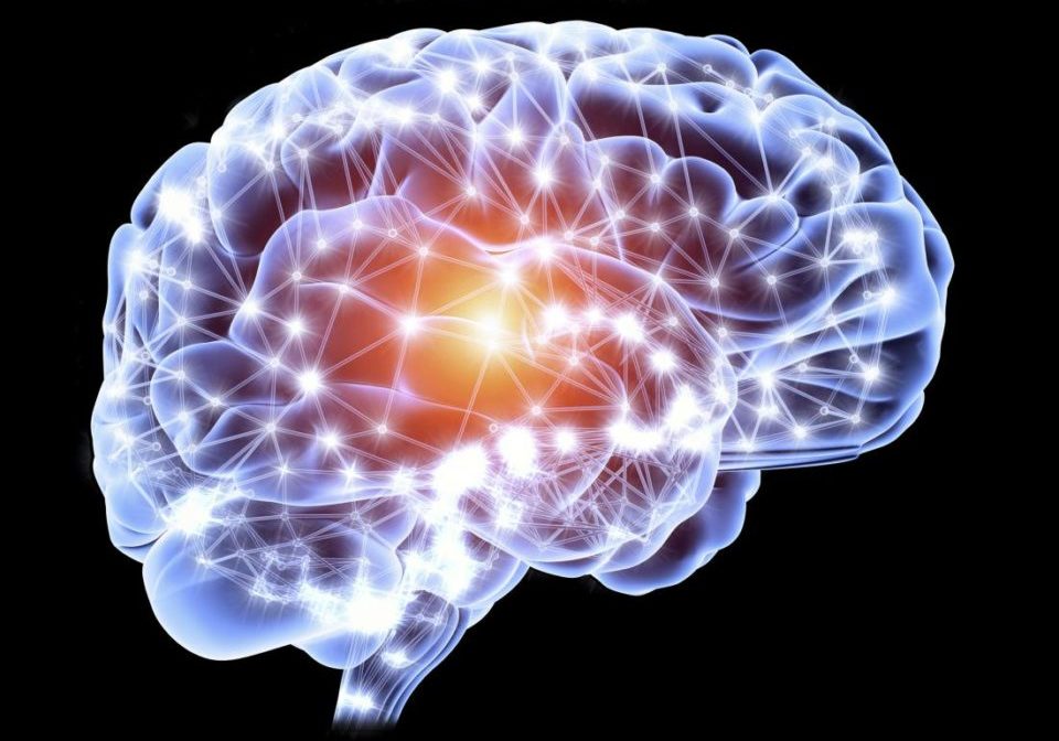
Individuals with ASD tend to disengage from social interactions when compared to people who are typically developing (TD). Scientists hypothesize that one reason for their disengagement may lie in the value, or lack thereof, their brains place on social exchanges. Neurobiological investigations have identified that a part of the brain called the ventral medial prefrontal cortex (vmPFC) is critical for encoding the value of different actions corresponding to distinct environmental cues and could be used to detect ASD.
“Right now, a two- to four-hour session by a qualified clinician is required to diagnose autism, and ultimately it is a subjective assessment based on their experience,” said the study’s principal investigator, Kenneth Kishida, Ph.D., assistant professor of physiology and pharmacology at Wake Forest School of Medicine, part of Wake Forest Baptist Health, in a press release.
The study comprised 40 participants (age range, 6-18 years old), of which 12 had ASD and 28 were TD. Participants were prompted to view images of four faces and four objects, which were placed on a projection screen and viewed through a mirror during fMRI scanning. Each image was categorized as either: favorite, pleasant, neutral, or unpleasant. The “favorite” images depicted each of the participants’ self-selected favorite person and thing, while the rest were chosen and designed from the International Affective Picture System (IAPS) database. Subsequently, each of the eight images were shown once for only for five seconds in a block that repeated six times. Following the completion of the 12- to 15-minute MRI scans, the participants were shown the same set of images and ranked in order from pleasant to unpleasant using a self-assessing sliding scale.
Able to Differentiate Between ASD and TD
Results of the study indicate that the vmPFC elicited a stronger response to favorite pictures in TD children, but not in children in ASD. The researchers were able to differentiate the ASD and TD cohorts after just 30 seconds of fMRI data capturing single stimulus images. “How the brain responded to these pictures is consistent with our hypothesis that the brains of children with autism do not encode the value of social exchange in the same way as typically developing children,” Dr. Kishida said.
Scientists succeed in testing potential #brain-based method to diagnose #autism https://t.co/8ObnkItoLg
— Medical Xpress (@medical_xpress) May 20, 2019
Overall, Dr. Kishida was pleased with the findings of this study, and looking ahead to the future said that “based on our study, we envision a test for autism in which a child could simply get into a scanner, be shown a set of pictures and within 30 seconds have an objective measurement that indicates if their brain responds normally to social stimulus and non-social stimuli.”
Scientists succeed in testing potential brain-based method to diagnose autism https://t.co/pCD8T27JvE #Psychology
— 🧠 Larry G. Maguire (@LarryGMaguire) May 20, 2019
Scientists succeed in testing potential brain-based method to diagnose autism https://t.co/p5lUAJYA4X pic.twitter.com/D4C3hQ5jxz
— Bioengineer.org (@bioengineerorg) May 20, 2019
Source: Biological Psychology, Wake Forest School of Medicine

 © 2025 Mashup Media, LLC, a Formedics Property. All Rights Reserved.