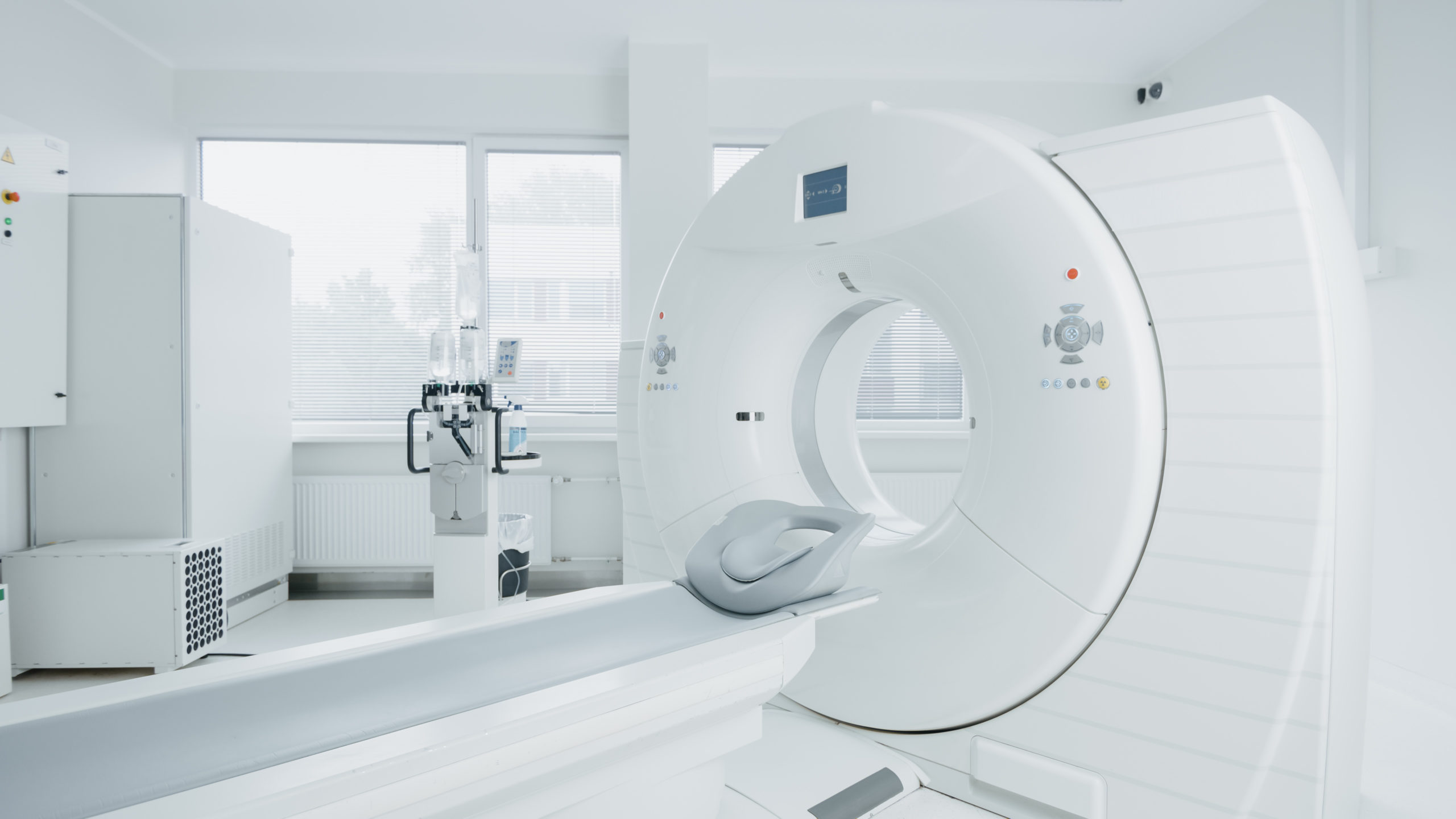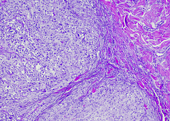
In a meta-analysis published in European Urology Oncology, researchers examined the diagnostic accuracy of prostate-specific membrane antigen positron emission tomography targeted biopsy (PSMA-PET-TB) for the detection of clinically significantly prostate cancer (csPCa) in biopsy samples. According to lead author, Tatsushi Kawada, and colleagues, PSMA-PET was a “promising imaging method” for detecting csPCa, and seemed to add additional value when combined with magnetic resonance imaging (MRI) detection.
The systematic literature review identified five prospective studies encompassing 497 patients which were published to the PubMed, Web of Science, and Scopus databases by December 2021. Two of the studies evaluated PSMA-PET-TB alone and the other three evaluated PSMA-PET-TB plus MRI-TB. The primary measures of the analysis were pooled sensitivity, specificity, positive predictive value (PPV), and negative predictive value (NPV).
Pooled Outcomes of PSMA-PET-TB With or Without MRI-TB
According to the report, PSMA-PET-TB alone showed the following pooled diagnostic values for csPCa detection:
- Sensitivity: 0.89 (95% CI, 0.85–0.93)
- Specificity: 0.56 (95% CI, 0.29–0.80)
- PPV: 0.69 (95% CI, 0.58–0.79)
- NPV: 0.78 (95% CI, 0.50–0.93)
Based on the three studies which evaluated PSMA-PET-TB plus MRI-TB, the authors calculated the following pooled estimates:
- Sensitivity: 0.91 (95% CI 0.77–0.97)
- Specificity: 0.64 (95% CI 0.40–0.82)
- PPV: 0.75 (95% CI 0.56–0.87)
- NPV: 0.85 (95% CI 0.62–0.95)
Additionally, in examining lesions with a Prostate Imaging-Reporting and Data System (PI-RADS) score of 3, the combined strategy showed a sensitivity, specificity, PPV, and NPV of 0.69, 0.73, 0.48, and 0.86, respectively.
The authors noted limitations including a low number and strength of reviewed studies which, despite promising pooled synthesis results, limits the borad implementation of PSMA-PET in clinical practice. Also, the included studies had different definitions for csPCa, criteria for positive imaging, and PSMA-PET ligands used.
Moreover, two studies only enrolled biopsy-naïve patients—with the majority from one of the two studies—while the others included naïve and repeated biopsy patients. Although the analysts didn’t identify significant differences between the subgroups, they noted they could not separately analyze repeat-biopsy only participants.
Taken together, the authors acknowledged heterogeneity and sample size biases may limit the generalizability of their PSMA-PET-TB findings. Nonetheless, they reported PSMA-PET-TB demonstrated promising diagnostic utility, and that it might add value to multiparametric MRI (mpMRI), particularly for PI-RADS 3 lesions.
For current practice, they advised that “PSMA-PET for diagnostic use should be considered in combination with mpMRI only for selected patients, while mpMRI before prostate biopsy and MRI-TB should be recommended.” They suggested PSMA-PET could allow for improved risk classification for suspected csPCa, and closed with a call for further studies “to standardize PSMA-PET recommendations for PCa detection.”
Related: Mechanisms of Testosterone in Prostate Cancer Progression


 © 2025 Mashup Media, LLC, a Formedics Property. All Rights Reserved.