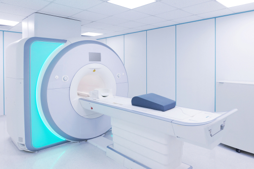
A noninvasive magnetic resonance imaging (MRI) technique can effectively evaluate bone marrow in patients with myelofibrosis (MF), according to a study presented at the 65th ASH Annual Meeting & Exposition.
Tanner Robison, PhD, and colleagues analyzed 66 study subjects (45 with MF, 15 with essential thrombocytosis or polycythemia vera, and 6 healthy controls) using 3 quantitative MRI sequences: proton density fat fraction for fat content, apparent diffusion coefficient for cellular effect on water mobility, and magnetization transfer for macromolecular structure and fiber deposition. They then linked MRI metrics to patient prognosis.
According to the findings, individual MRI metrics correlated significantly across anatomic sites (rpearson from 0.57 to 0.89; P<.01), with notable heterogeneity observed both within and among different regions. Dynamic International Prognostic Scoring System risk groups had limited correlation with MRI metrics. Moreover, a logistic regression model demonstrated that quantitative MRI metrics were associated with the severity of fibrosis in the bone marrow (test set performance: accuracy, 84.6%; sensitivity to predict overt fibrosis, 88.9%; specificity, 75.0%; area under the receiver operator curve, 0.94).
“Quantitative bone marrow MRI reliably captures relevant metrics of MF bone marrow, including cellularity and fat replacement by fibrosis, and may be useful in clinical drug trials and patient management for MF,” the researchers concluded.
Reference
Robinson T, Levinson A, Lee W, et al. MRI reliably captures bone marrow metrics in myelofibrosis. Abstract #4556. Presented at the 65th ASH Annual Meeting & Exposition; December 9-12, 2023; San Diego, California.


 © 2025 Mashup Media, LLC, a Formedics Property. All Rights Reserved.