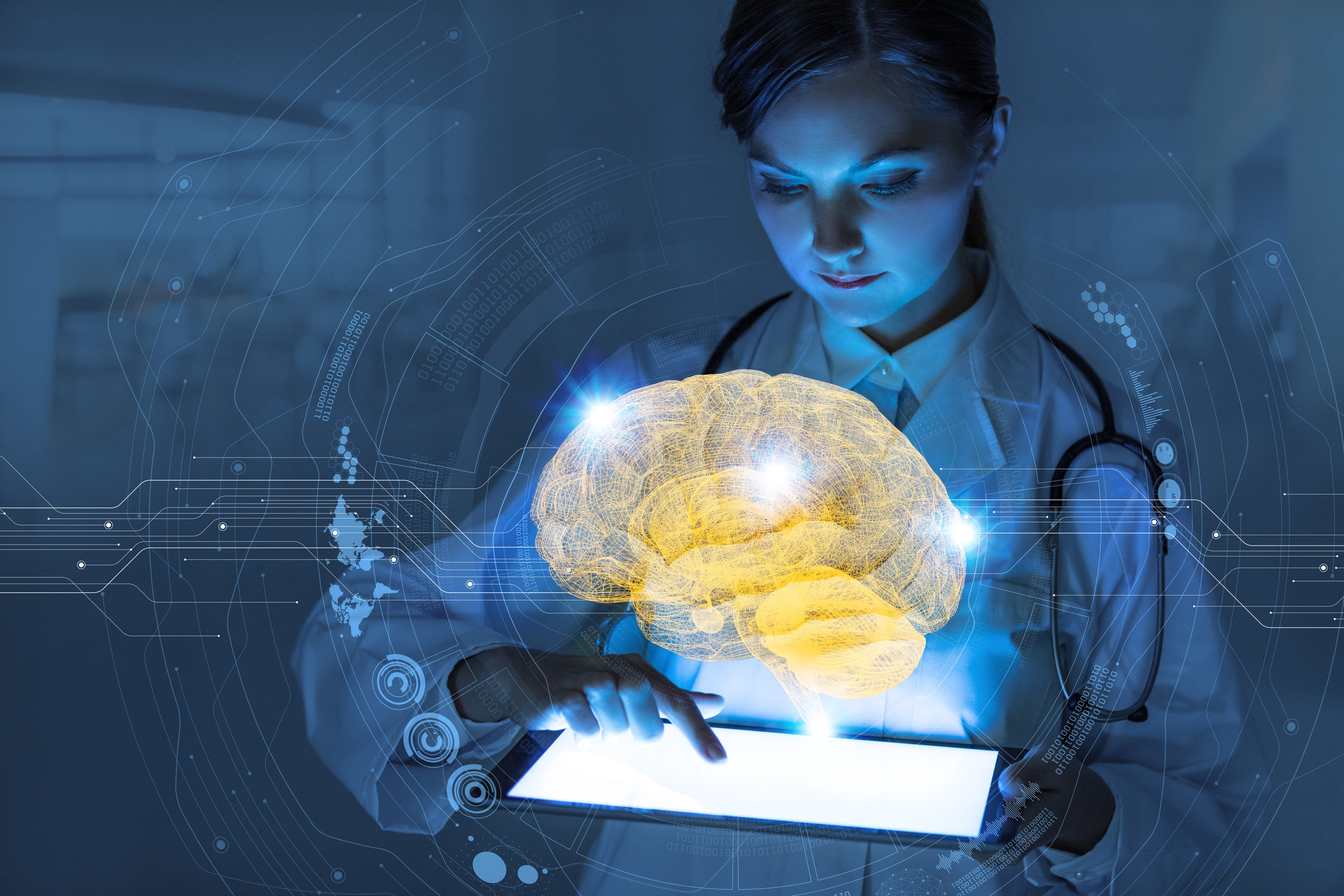
A research team from the University of Malaga (UMA)’s ICAI Group -Computational Intelligence and Image Analysis- have recently created an artificial intelligence (AI) platform that can improve magnetic resonance imaging (MRI) of the brain. This technology leverages a deep learning neural network to increase the quality of such images without distorting the patients’ neurological structures. This specific type of AI is designed to mimic the human brain’s learning process and was reported in the journal Neurocomputing.
The MRI process is far from perfect, and can occasionally generate poor quality images that are of little help to the physician. To combat this, researchers have begun creating “super-resolution” images, or images with increased resolution based on examples that identify the differences between low and high resolution images. This can be achieved through a deep learning neural network, which can estimate this association with high accuracy.
“Deep learning is based on very large neural networks, and so is its capacity to learn, reaching the complexity and abstraction of a brain”, explained the study’s lead author, Karl Thurnhofer. This technique also allows the identification to be performed autonomously, with no human intervention, as per Thurnhofer. This task carried out by the AI system is one that the human eye is incapable of performing.
Thurnhofer’s work has potentially significant implications for the field of radiology, being that this UMA team’s algorithm is capable of generating results of higher quality in a shorter amount of time, presenting benefits to both the patient, physician, and the healthcare industry.
“So far, the acquisition of quality brain images has depended on the time the patient remained immobilized in the scanner; with our method, image processing is carried out later on the computer,” said Thurnhofer.
This deep learning solution has the potential to allow specialists to identify brain pathologies tied to cancer, physical injury, language or motor disorders, with greater accuracy. In addition, these thinner images will improve the diagnostic capability of these images as well, minimizing the need for additional imaging procedures to be done.
UMA’s ICAI Group is led by Professor Ezequiel López who is also a co-author of this study. Enrique Domínguez and Rafael Luque, Professors of the Department of Computer Science and Programming Languages also participated in this study, as did as researcher Núria Roé-Vellvé.
“The results obtained on different MR images show a considerable improvement both in the restored image and in the residual image without an excessive increase in computing time,” the authors of the study concluded.
Artificial intelligence to improve resolution of brain magnetic resonance imaging https://t.co/9AT5wohh8k pic.twitter.com/fipv3MhTrX
— Paul McCullough (@ThePostsynaptic) January 19, 2020


 © 2025 Mashup Media, LLC, a Formedics Property. All Rights Reserved.