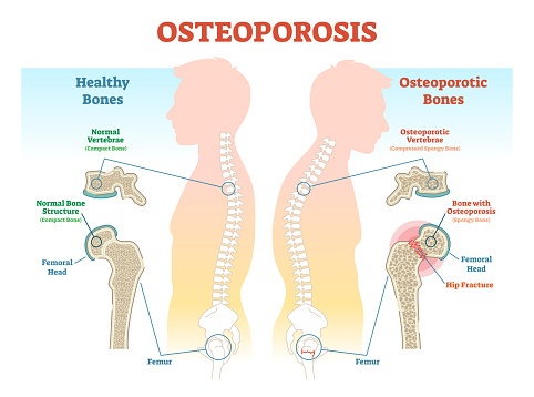
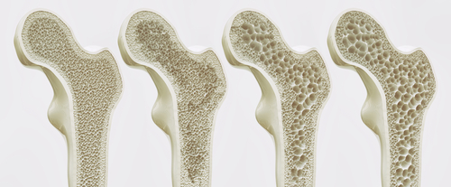
Among patients who received a quantitative computed tomography (QCT) scan of the lumbar spine prior to undergoing spinal fusion surgery, many were found have low bone density, and in almost half those cases, were diagnosed with osteoporosis, or its precursor, osteopenia, according a recent study at the Hospital for Special Surgery (HSS) in New York City.
The research, titled, “Prevalence of Osteopenia and Osteoporosis Diagnosed by Quantitative Computed Tomography in 296 Consecutive Lumbar Fusion Patients,” was presented yesterday at the American Academy of Orthopaedic Surgeons Annual Meeting in Las Vegas.
Spinal fusion surgery is very common, with 400,000 to 500,000 such procedures performed annually in the US. Of the total number of spinal fusions completed, two-thirds occur in the lumbar region. The standard test employed to measure bone strength and detect low bone density is dual energy X-ray absorptiometry (DXA or DEXA), a type of flat x-ray that reads bone material content. QCT measures bone mineral density with a CT scanner, resulting in a 3D image.
The Advantages of QCT
“The literature reporting QCT-based lumbar spine bone density is scarce, and we believe our study is the first of its kind,” said Dr. Alexander Hughes, an orthopedic surgeon specializing in spine surgery at HSS and senior study investigator, in a press release. “We believe that QCT is more effective in screening patients because the DXA scan can overestimate bone density in the spine due to certain bone changes, a patient’s weight or physique, and other factors.”
In this study, the researchers recruited 296 patients (age range, 21 to 89; mean age, 63; 55% female) undergoing lumbar spine fusion for either a degenerative condition or spinal instability. All patients had preoperative QCT scans.
CT scan prior to spine fusion finds almost half of patients had undiagnosed osteoporosis https://t.co/y0Lpth84iK #Imaging
— IS&T (@ImagingOrg) March 12, 2019
High Percentage of Low Bone Density
Researchers used the American College of Radiology criteria to uncover that 44% of patients had osteopenia, and 15% had osteoporosis, while 41% had normal bone density. The researchers reported no discernible differences in prevalence between gender or race, but did indicate that patients over 50 were far more likely to incur low density than younger patients. Moreover, among patients under 50, while none were found to have osteoporosis, 17% were discovered to have osteopenia. Furthermore, within a subgroup of 212 patients with no previous history of low bone density, 39% were diagnosed with osteopenia and 10% had osteoporosis.
“Spine surgeons should be aware of the high prevalence of abnormal bone mineral density in lumbar spine patients and the possibility that those without a previous diagnosis may have osteopenia or osteoporosis,” said Dr. Hughes. “Diagnosing this prior to spine fusion could lead to a change in surgical planning and treatment, and we believe this would improve outcomes.”
CT scan prior to #spine fusion finds almost half of patients had undiagnosed #osteoporosis @hspecialsurgery https://t.co/rhTv5WlSQ9
— Medical Xpress (@physorg_health) March 12, 2019
https://twitter.com/Social_Media_b8/status/1105506385104654337


 © 2025 Mashup Media, LLC, a Formedics Property. All Rights Reserved.