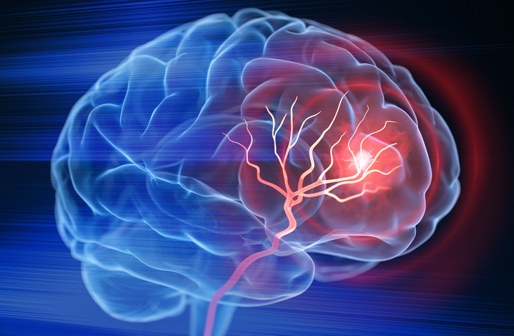
A novel imaging technique added to MRI allows clinicians to better tailor rehabilitation for stroke patients, according to the findings of a study that appeared in Neurology.
The analysis, conducted by Georgetown University Medical Center in concert with MedStar Health and the National Institutes of Health, assessed a technique called diffusion tensor-based morphometry (DTBM). Compared with conventional TBM techniques, DTBM provides a more accurate look at volume changes in white matter structures in the brain. Essentially, this new method allows clinicians to observe white matter tracts leading to the limbs more clearly, enabling them to render a more accurate rehabilitation path.
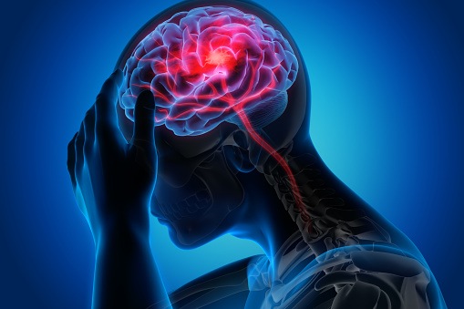
“A white matter tract called the corticospinal tract provides the main wiring that goes from your brain down to your spinal cord to help power your arms and legs,” explains study leader, Matthew A. Edwardson, MD, an associate professor of neurology at Georgetown University School of Medicine and a vascular neurologist and a member of the stroke team at MedStar Georgetown University Hospital. “If those cables are severed or atrophied, the person is not going to be able to regain meaningful use of their arms and legs – they wouldn’t have much motor strength.”
In this study, researchers evaluated 21 participants with anterior circulation stroke. The findings of the analysis showed that DTBM can detect volume loss in the corticospinal tract (CST) in the first 30 days following stroke. “These volumetric changes may provide complementary information to FA in characterizing white matter loss after stroke,” the researchers concluded.
Source:
Research supports move toward better tailoring stroke rehabilitation. News release. Georgetown University Medical Center. March 3, 2025. Accessed March 4, 2025. https://www.eurekalert.org/news-releases/1075487

 © 2025 Mashup Media, LLC, a Formedics Property. All Rights Reserved.