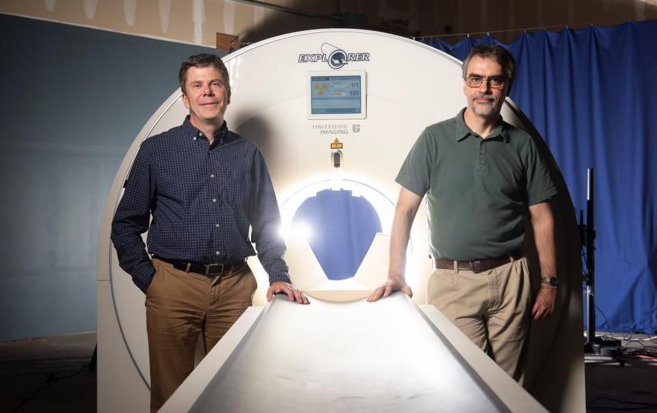
EXPLORER, the 3D medical imaging system that can scan the whole body at once, has recently produced its first scans. Created by UC Davis researchers Simon Cherry and Ramsey Badawi, the scanner combines positron emission tomography (PET) and x-ray computed tomography (CT) technologies to image the entire human body. Coupling these two imaging methods and capturing radiation much more effectively than other scanning systems, EXPLORER can generate images much faster than other scanning methods. Over time, this system can also create videos tracking specially marked drugs as they travel throughout the body.
The scientists claim that EXPLORER will have many applications in the medical field, including diagnostic improvements, tracking disease progression, and researching novel drug therapies. Badawi and Cherry devised the concept of a full body scanner 13 years ago, receiving a $1.5 million grant from the National Cancer Institute in 2011. This grant allowed the two to establish a team of researchers and collaborators to bring the idea to life. Their funds were greatly bolstered in 2015 when the NIH awarded them a $15.5 million grant, allowing Badawi and Cherry to enlist a commercial partner to build the first EXPLORER scanner.
The first images generated by EXPLORER will be shown at the upcoming Radiological Society of North America meeting on November 24th in Chicago.
This scanner was created in partnership with United Imaging Healthcare (UIH), a Shenghai-based company who built the system with its latest technology platform. Eventually, UIH will be manufacturing the devices for the consumers in the healthcare market.
“While I had imagined what the images would look like for years, nothing prepared me for the incredible detail we could see on that first scan,” said Cherry, professor in the Department of Biomedical Engineering at UC Davis. “While there is still a lot of careful analysis to do, I think we already know that EXPLORER is delivering roughly what we had promised.”
Cherry claims he expects EXPLORER to have a heavy impact on patient care and research, being that it produces PET scans that are of higher-quality than others previously available. The scanner also yields results up to 40 times faster than current PET scans, imaging the whole body in less than 30 seconds.
Badawi, functioning chief of Nuclear Medicine at UC Davis Health and vice research chair in the Radiology Department, was astounded by the resolution of the first images. He stated that, “the level of detail was astonishing, especially once we got the reconstruction method a bit more optimized.”
“We could see features that you just don’t see on regular PET scans. And the dynamic sequence showing the radiotracer moving around the body in three dimensions over time was, frankly, mind-blowing. There is no other device that can obtain data like this in humans, so this is truly novel,” Badawi added.
The video below shows EXPLORER’s ability to track a glucose injection through a vein in the leg.
“I don’t think it will be long before we see at a number of EXPLORER systems around the world,” stated Cherry. “But that depends on demonstrating the benefits of the system, both clinically and for research. Now, our focus turns to planning the studies that will demonstrate how EXPLORER will benefit our patients and contribute to our knowledge of the whole human body in health and disease.”
Sources: EurekAlert, CNET


 © 2025 Mashup Media, LLC, a Formedics Property. All Rights Reserved.