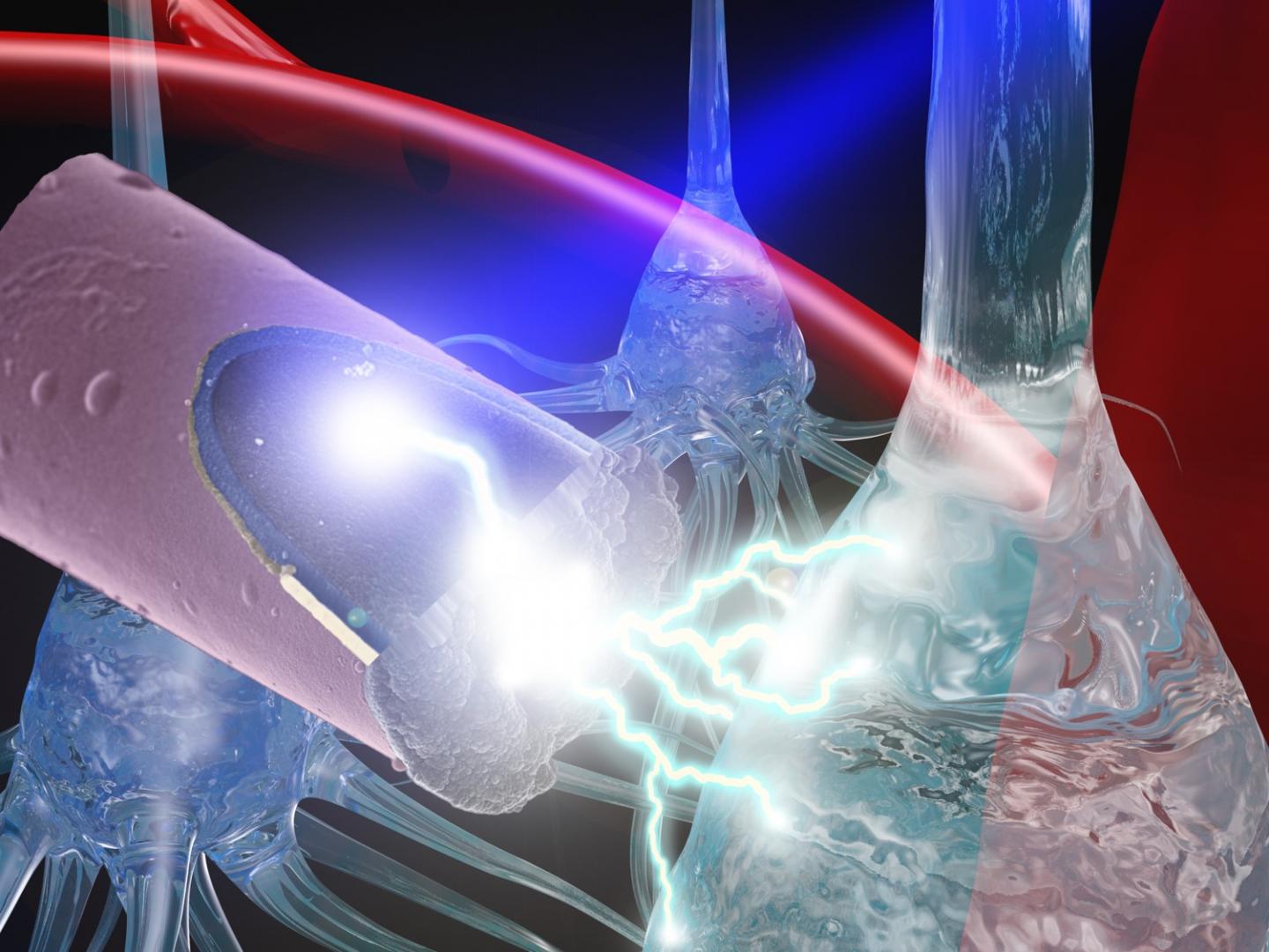
A team of bionengineers from the University of Pittsburgh have recently developed a neural stimulation technique that may be safer than current methods. These electrode stimulation techniques are used in treating patients with spinal cord injury, sensory deficits, strokes, and other neurological disorders such as Parkinson’s disease. Despite their therapeutic benefits, these implanted devices commonly degrade over time and create scarring in neural tissue. Led by Takashi D. Y. Kozai, these Pitt researchers made use of a light activated, untethered ultrasmall electrode that may cause less damage than current electrodes.
“Typically with neural stimulation, in order to maintain the connection between mind and machine, there is a transcutaneous cable from the implanted electrode inside of the brain to a controller outside of the body,” stated Kozai, assistant professor of bioengineering in Pitt’s Swanson School of Engineering. “Movement of the brain or this tether leads to inflammation, scarring, and other negative side effects. We hope to reduce some of the damage by replacing this large cable with long wavelength light and an ultrasmall, untethered electrode.”
The first author of the study, Kaylene Stocking, is a senior bioengineering and computer engineering student, works with Kozai’s Bionic Lab to investigate how to increase longevity of neural implant technologies. “Intracortical neural stimulation with untethered, ultrasmall carbon fiber electrodes mediated by the photoelectric effect,” Kozai said.
READ MORE: UTSA Integrating Virtual Reality (VR) Into Student Education
The photoelectric effect refers to the phenomenon occurring when a photon contacts an object and causes local changes in electrical potential. Kozai and his team found this techniques advantages when conducting other imaging research projects. Based on previous findings of this effect published in 1905 by Einstein, the researchers expected to observe electrical photocurrents only at ultraviolet wavelengths, but their findings surprised them.
“When the photoelectric effect contaminated our electrophysiological recording while imaging with a near-infrared laser (low energy photons), we were a little surprised,” explained Kozai. “It turned out that the original equation had to be modified in order to explain this outcome. We tried numerous strategies to eliminate this photoelectric artifact but were unsuccessful in each attempt, so we turned the ‘bug’ into a ‘feature.’”
READ MORE: How Companies Are Integrating Voice Recognition Into Medicine
Using a carbon fiber implant that is 7-8 microns in diameter (slightly smaller than a neuron), Stocking was able to simulate their technique on a ‘phantom’ brain using a two-photon microscope. She used this test to analyze the effects of this method to see if the electrical potential stimulated the cells in a fashion similar to neural stimulation.
“Our group decided to use this feature of the photoelectric effect to our advantage in neural stimulation,” said Stocking. “We used the change in electrical potential with a near-infrared laser to activate an untethered electrode in the brain. We discovered that photostimulation is effective. Temperature increases were not significant, which lowers the chance of heat damage, and activated cells were closer to the electrode than in electrical stimulation under similar conditions, which indicates increased spatial precision.”
Kozai noted that have had several critics who did not trust the mathematical modifications made to Einstein’s photoelectric equation, but that he and his team have faith in the technique and have filed a patent application. “This is a testament to Kaylene’s hard work and diligence to take a theory and turn it into a well-controlled validation of the technology,” he concluded.
Pitt bioengineers create ultrasmall, light-activated electrode for neural stimulation https://t.co/lS8FggTUkx pic.twitter.com/rmgcJXFS5p
— YTLC (@YTLC1) February 15, 2019
Sources: EurekAlert, IEEE Xplore


 © 2025 Mashup Media, LLC, a Formedics Property. All Rights Reserved.