Resistance to targeted therapies continues to pose a significant challenge in treating metastatic melanoma. The tumor microenvironment (TME) plays a crucial role in cancer progression and the development of drug resistance. However, the advancement of such treatments has been hindered by the absence of human in vitro 3D models that accurately replicate the complex interactions between melanoma and the TME.
To overcome this limitation, researchers have developed integrated skin-TME-melanoma organoids (mTMEOs), which incorporate essential components of the melanoma TME. These organoids offer a realistic in vitro platform for studying TME-driven tumor progression and for evaluating potential therapeutic interventions. The results will be presented at the American Society of Clinical Oncology 2025 Annual Meeting.
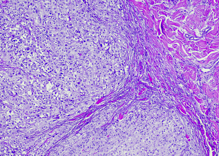
Primary human keratinocytes, melanocytes, fibroblasts, and pre-adipocytes were co-aggregated with GFP-labeled melanoma cells (SK-MEL-28 or A375) to create melanoma-skin organoids (mSOs), which successfully recapitulated key aspects of melanoma tissue architecture, as confirmed by immunostaining. To construct the melanoma tumor microenvironment organoids (mTMEOs), the mSOs were embedded in collagen gels containing fibroblasts to mimic the dermal stroma.
These mTMEOs were imaged every other day using the Vireo Multi-Camera Array Microscope (MCAM) under fluorescence. A custom ImageJ-based analysis pipeline was developed to track the GFP-labeled melanoma cells within the collagen matrix, enabling quantification of the number, size, and migratory distance of invading melanoma cell clusters.
The mSOs successfully replicated key structural features of human skin, including a stratified epidermis and a dermal-hypodermal core, as well as melanoma-specific characteristics such as epidermal spread, atypical melanocytes, and outward cell migration. In mTMEOs, a transition from radial to vertical growth was observed, enabling melanoma invasion into the dermis-like stromal matrix. Fluorescence imaging with the Vireo MCAM was used to create an Euclidean distance map, allowing quantification of melanoma migration from the nearest mSO.
Analysis revealed that while the presence of stromal fibroblasts in the collagen gels did not significantly affect invasion rates, it did promote greater migration distances and resulted in fewer but larger melanoma cell clusters compared with fibroblast-free gels. These results are consistent with existing studies suggesting that although dermal invasion may be initiated through interactions with various cell types, stromal fibroblasts may facilitate melanoma progression by promoting cell dispersal and favoring collective migration—a behavior linked to a more aggressive metastatic potential.
“Our novel mTMEO model replicates melanoma TME anatomical and pathological features, which can be altered by modifying individual organoid’s components. mTMEOs and the subsequent analysis workflow provide a versatile and tunable platform for studying TME-driven mechanisms of tumor progression and for drug development. By incorporating patient-derived cells, these constructs have the potential to serve as a personalized drug screening tool to improve patient outcomes,” the researchers concluded.
References
Nomdedeu-Sancho G, et al. 2025 ASCO Annual Meeting. Chicago, Illinois. May 30 to June 3, 2025. https://meetings.asco.org/abstracts-presentations/246658


 © 2025 Mashup Media, LLC, a Formedics Property. All Rights Reserved.