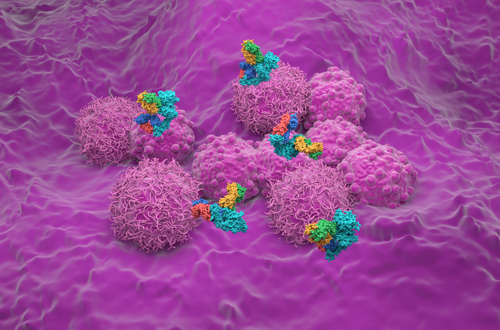
HER2-low breast cancer has emerged as a pivotal subtype in oncology following the promising outcomes of the DESTINY-Breast04 (DB-04) and DESTINY-Breast06 (DB-06) trials. For physicians managing metastatic breast cancer, distinguishing HER2-low from HER2-null is now clinically meaningful because patients with HER2-low tumors are eligible for trastuzumab deruxtecan (T-DXd) therapy. Yet this advancement poses new challenges for pathologists because traditional immunohistochemistry (IHC) scoring systems were not designed to reliably discern these subtle differences. A new Australian-led study provides a validated, HER2-low–focused IHC scoring system that enhances diagnostic consistency across expert pathologists.
Historically, HER2 IHC scores of 0 and 1+ were deemed clinically inconsequential, resulting in limited training or attention to low-expressing tumors. However, with T-DXd now FDA approved for HER2-low tumors, accurate stratification is vital. “Making the subtle distinctions in IHC staining patterns of various HER2-negative cancers presents a new and urgent diagnostic challenge,” the authors write. Compounding the issue is the lack of reference standards for HER2-low breast cancer, absence of confirmatory tests such as in situ hybridization, and variable interpretation of weak membrane staining. Retrospective audits in the US and Denmark revealed alarming interobserver variability, casting doubt on the reliability of existing methods.
Clinical Guidelines and Consensus
The study builds on the 2018 ASCO-CAP HER2 testing guidelines, expanding them with HER2-low–specific conventions. These include detailed instructions on slide evaluation, emphasizing the need to inspect all tumor regions at 40× magnification before assigning a score of 0. Unlike HER2-positive cancers, “HER2-low cancers often exhibit only partial, faint membrane staining in just over 10% of tumor cells,” necessitating a refined evaluation protocol. The authors’ guidelines also caution against misinterpretation of cytoplasmic or artifactual staining, which can falsely elevate HER2 scores.
One major barrier is the pathologist’s learning curve in differentiating HER2 0, 1+, ultralow (UL), and null types. To address this issue, the research team validated their scoring system using a new dataset of 64 digitized HER2-negative core biopsy specimens, ensuring preanalytical integrity and balanced case distribution. Nine expert breast pathologists from 5 Australian states scored the slides independently after a 5-month washout period from the initial study. By defining a “majority opinion” as the consensus standard and emphasizing training through a dedicated scoring flowchart, the team improved interobserver concordance.
Guidelines for Management
For clinicians, accurate HER2-low classification determines eligibility for T-DXd. According to the DESTINY-Breast04 trial, patients with IHC scores of 1+ or 2+ (without HER2 amplification) showed improved progression-free and overall survival with T-DXd compared with chemotherapy of physician’s choice. In the newer DB-06 trial, even patients with HER2-ultralow expression showed encouraging trends, although statistical significance is pending. Pathologists must therefore be equipped to identify HER2-low cases with precision, ensuring that eligible patients are not excluded from effective therapies.
Although the study focuses on pathology, patient-facing clinicians play a vital role in explaining HER2 status and its evolving implications. As testing criteria shift, patients may have questions about changes in eligibility or treatment plans. Clear, empathetic communication about what HER2-low means and why their tumor may now qualify for targeted therapy is essential for shared decision-making and treatment adherence.
Conclusion
The validation of a HER2-low–focused IHC scoring system represents a significant advancement in diagnostic pathology. This study demonstrates that with targeted training and standardized guidelines, pathologists can achieve high concordance and accuracy, even in the nuanced realm of HER2-low assessment. As new therapies like T-DXd expand their indications, scalable solutions such as algorithm-assisted scoring and national quality assurance modules will be key. Ultimately, the model set forth by this Australian team offers a roadmap for global pathology communities aiming to meet the evolving demands of precision oncology.
BIOBOX
Authors
- Gelareh Farshid, MBBS, PhD, Discipline of Medicine, Adelaide University and SA Pathology, Royal Adelaide Hospital
- Jane Armes, Sullivan Nicolaides Pathology, Queensland
- Benjamin Dessauvagie, Clinipath Pathology and Medical School, Perth
- Amardeep Gilhotra, SA Pathology, Royal Adelaide Hospital
- Beena Kumar, Monash Health Pathology, Melbourne
- Hema Mahajan, St George Hospital, NSW
- Ewan Millar, NSW Health Pathology
- Nirmala Pathmanathan, Westmead Breast Cancer Institute
- Cameron Snell, Peter MacCallum Cancer Centre, Melbourne
Readings and Resources
- Modi S, Jacot W, Yamashita T, et al. Trastuzumab deruxtecan in previously treated HER2-low advanced breast cancer. N Engl J Med. 2022;387(1):9-20. doi:10.1056/NEJMoa2203690
- Curigliano G, Hu X, Dent RA, et al. Trastuzumab deruxtecan (T-DXd) vs physician’s choice of chemotherapy (TPC) in patients (pts) with hormone receptor-positive (HR+), human epidermal growth factor receptor 2 (HER2)-low or HER2-ultralow metastatic breast cancer (mBC) with prior endocrine therapy (ET): primary results from DESTINY-Breast06 (DB-06). J Clin Oncol. 2024;42:LBA1000. https://doi.org/10.1200/JCO.2024.42.17_suppl.LBA1000
- Farshid G, Armes J, Dessauvagie B, et al. Independent validation of a HER2-low focused immunohistochemistry scoring system for enhanced pathologist precision and consistency. Mod Pathol. 2025;38(4):100693. doi:10.1016/j.modpat.2024.100693
- Allison KH, Wolff AC. ERBB2-low breast cancer-is it a fact or fiction, and do we have the right assay? JAMA Oncol. 2022;8(4):610-611. doi:10.1001/jamaoncol.2021.7082
- Wolff AC, Hammond MEH, Allison KH, et al. Human epidermal growth factor receptor 2 testing in breast cancer: American Society of Clinical Oncology/College of American Pathologists Clinical Practice guideline focused update. Arch Pathol Lab Med. 2018;142(11):1364-1382. doi:10.5858/arpa.2018-0902-SA


 © 2025 Mashup Media, LLC, a Formedics Property. All Rights Reserved.