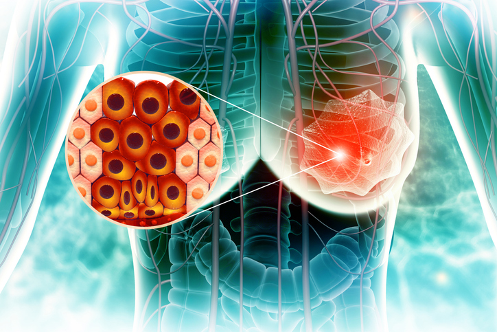
The classification of HER2-low breast cancer has gained significance with the advent of HER2-targeted therapies like trastuzumab deruxtecan (T-DXd). While HER2-low was previously considered clinically irrelevant, it is now recognized as a distinct therapeutic category with implications for treatment eligibility and prognosis. This has prompted oncologists and pathologists to reconsider diagnostic and prognostic approaches. A recent study by Giovanni De Rosa, MD, PhD, Department of Advanced Biomedical Sciences, University of Naples Federico II, Italy, offers cytological insights into HER2-low breast carcinoma, focusing on metastatic cases and analyzing how proliferation markers and survival outcomes compare to HER2-positive and HER2-negative cancers.
HER2-low breast cancer is defined as tumors with immunohistochemistry (IHC) scores of 1+ or 2+ with negative in situ hybridization (ISH). However, this definition introduces diagnostic ambiguity, as HER2-low is not a biologically distinct entity. “HER2-low identification relies heavily on the precision of cytological and histopathological evaluations,” the study notes. The lack of clear guidelines for HER2-low categorization, especially in metastatic settings, complicates treatment planning. Additionally, the biological behavior of HER2-low tumors remains underexplored, particularly regarding proliferative activity and survival outcomes.
Clinical Guidelines and Consensus
Current clinical guidelines from ASCO and CAP continue to evolve considering recent findings. While they provide criteria for HER2 testing, they do not yet offer comprehensive guidance specific to HER2-low cytological features in metastatic contexts. Dr. De Rosa’s study provides needed clarity by analyzing fine needle aspiration (FNA) samples from metastatic sites, thus aligning cytological observations with IHC-defined HER2 expression. The research highlights a trend toward higher proliferation indices in HER2-low tumors compared to HER2-negative but lower than HER2-positive cases.
A major limitation in evaluating HER2-low carcinomas in metastatic settings is the underuse of cytology, which is often regarded as inferior to histology. “Cytological samples, when adequately prepared and interpreted, can provide reliable HER2 and Ki-67 evaluations,” the authors emphasized. However, many institutions still underutilize cytological techniques for biomarker evaluation due to perceived variability and lack of standardized preparation. To overcome these challenges, the study advocates for the implementation of uniform cytological staining protocols, enhanced training, and inter-laboratory collaboration.
Additionally, the integration of proliferation markers like Ki-67 into the routine evaluation of HER2-low tumors remains inconsistent. The study underscores the prognostic value of Ki-67, particularly in distinguishing HER2-low from HER2-null tumors. Encouraging the adoption of this marker in both cytological and histological contexts could support more individualized treatment decisions. As new HER2-directed therapies emerge, the need for comprehensive, cytology-inclusive diagnostic strategies becomes more urgent.
Guidelines for Management
While HER2-low cancers do not qualify for trastuzumab or pertuzumab, they are now candidates for T-DXd in metastatic settings. This shift in treatment criteria has prompted a reassessment of HER2 categorization and raised the clinical stakes for accurate classification. In Dr. De Rosa’s study, HER2-low tumors exhibited intermediate Ki-67 levels and survival outcomes between HER2-positive and HER2-negative groups. These findings suggest that HER2-low status may serve as both a predictive and prognostic biomarker, potentially guiding therapeutic intensity, clinical trial enrollment, and post-treatment surveillance strategies.
For clinicians, this means that a nuanced understanding of HER2 expression—beyond the binary positive/negative system—is critical. Dr. De Rosa’s findings support the inclusion of cytological HER2 testing in metastatic workups, particularly when tissue biopsies are not feasible. Standardizing HER2-low reporting language and integrating cytological data into multidisciplinary tumor boards may also improve treatment planning across institutions.
As classifications evolve, oncologists must communicate to patients that HER2-low status, once overlooked, now plays a pivotal role in determining treatment options. Explaining the nuances of HER2 expression can help patients make informed decisions about targeted therapies and clinical trials. In metastatic settings, where treatment decisions are time-sensitive, clear dialogue around HER2 status is essential.
Advancing Personalized Care in HER2-Low Breast Cancer
HER2-low breast cancer represents a new frontier in personalized oncology. The study by Dr. De Rosa emphasizes the value of cytological assessments in metastatic settings, highlighting their reliability in evaluating HER2 status and proliferation. With HER2-low now influencing treatment eligibility and outcomes, integrating cytological markers like Ki-67 into routine evaluation can enhance precision. Future guidelines must expand to include cytology-specific standards, especially for metastatic disease, to ensure patients receive optimal, targeted care.
BIOBOX
Authors
- Giovanni De Rosa, MD, PhD, Department of Advanced Biomedical Sciences, University of Naples Federico II, Naples, Italy
- Federica Zito Marino, Maria Vittoria Di Crescenzo, Sara Caiazza, Maddalena Raia, Giuseppina Liguori, Ilaria Aquino, Stefania Cantile, and Gerardo Botti, Istituto Nazionale Tumori IRCCS “Fondazione G. Pascale,” Naples, Italy
Reference
- Hrizat AS, Doxzon KA, O’Neill R, Post RP, Brachtel EF. Evaluation of HER2-Low breast carcinoma in metastatic settings: a cytological approach to proliferation and survival. J Am Soc Cytopathol. Published online January 6, 2025. doi:10.1016/j.jasc.2025.01.001


 © 2025 Mashup Media, LLC, a Formedics Property. All Rights Reserved.