
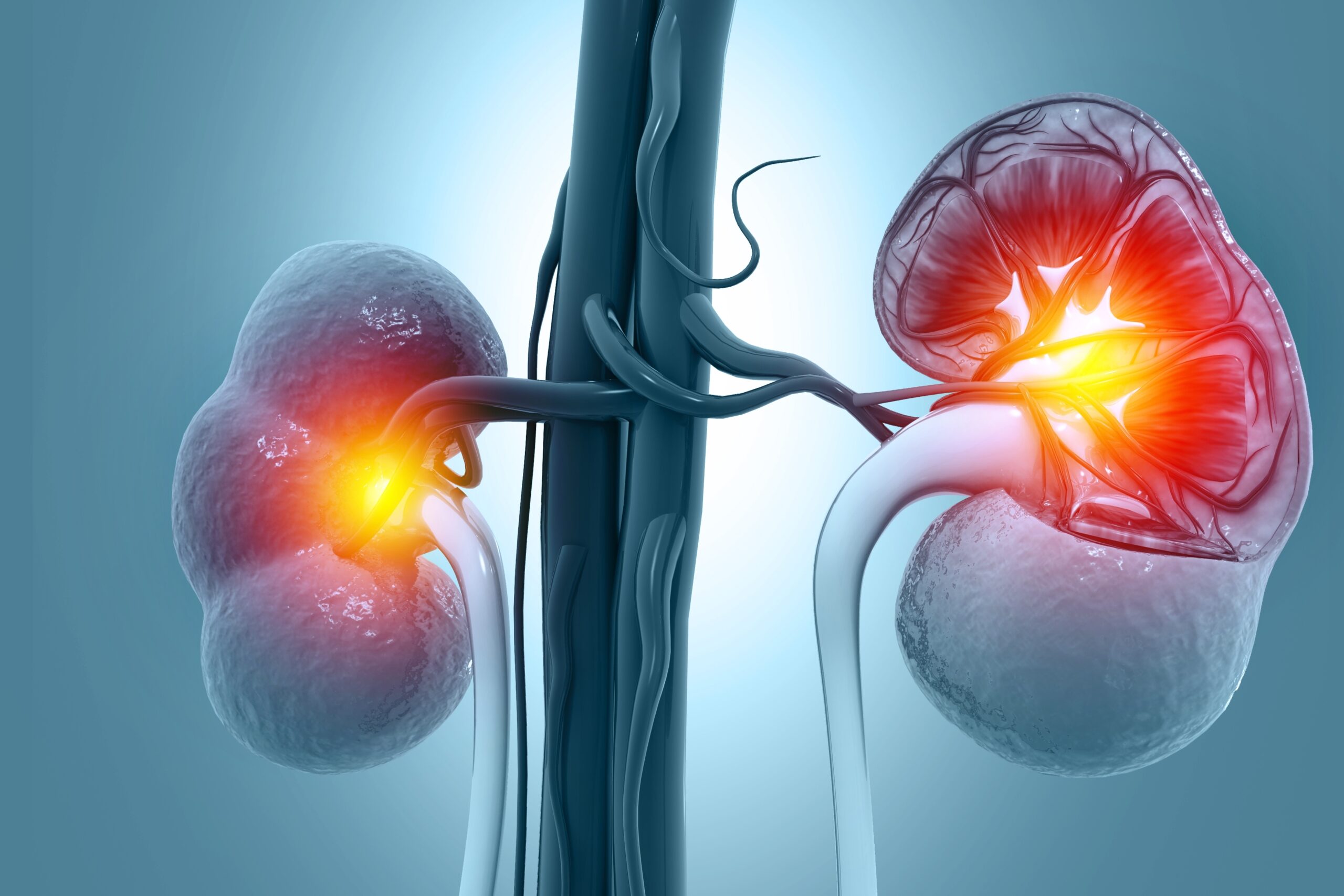
Huan Zhou, MD, and colleagues conducted a study exploring the efficacy of diffuse magnetic resonance imaging (MRI) to identify clinicopathological changes in patients with immunoglobulin A nephropathy (IgAN). Their findings were published in the British Journal of Radiology.
The study included 46 patients with IgAN and 27 healthy volunteers. The participants with IgAN were divided into three groups: group 1 (eGFR ≥90 mL/min/1.73 m2), group 2 (eGFR 60-90 mL/min/1.73 m2), and group 3 (eGFR <60 mL/min/1.73 m2). The researchers performed intravoxel incoherent motion diffusion-weighted imaging (IVIM-DWI) and diffusion tensor imaging (DTI) with 3.0 T magnetic resonance. They also collected and analyzed diffuse MRI, clinical, and pathological indicators.
The apparent diffusion coefficient, diffusion coefficient, perfusion fraction, and fractional anisotropy (FA) differed significantly among the IgAN subgroups and controls. These factors positively correlated with eGFR and negatively correlated with creatinine. They inversely correlated with glomerular sclerosis, interstitial fibrosis, and tubular atrophy (all P<.05). These parameters had significantly high area under the curve (AUC) for differentiating IgAN patients from controls. FA demonstrated the highest AUC for identifying group 1 IgAN patients versus volunteers.
The authors summarized, “DTI and IVIM-DWI had the advantage of evaluating clinical and pathological changes in IgAN patients. DTI was superior at distinguishing early IgAN patients and might be a noninvasive marker for screening early IgAN patients from healthy individuals.”
Source: British Journal of Radiology


 © 2025 Mashup Media, LLC, a Formedics Property. All Rights Reserved.