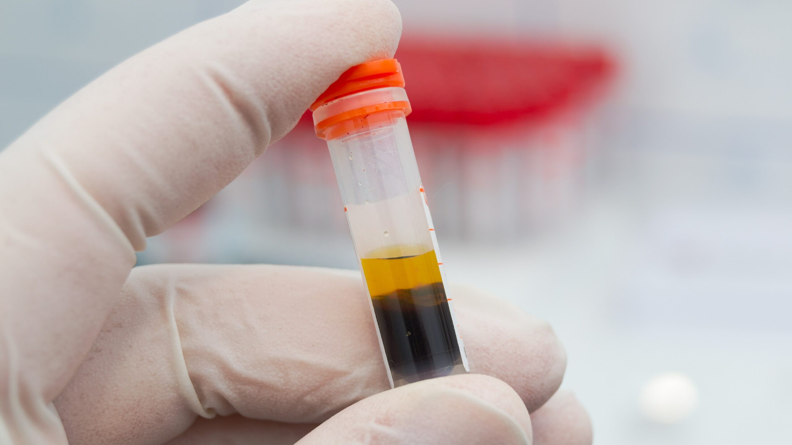
Ultrasonography examination enables detection of hyperechoic crystal deposits in the kidney medulla of patients with gout. Patients with chronic kidney disease (CKD) commonly experience hyperuricemia. There are few data available on whether hyperechoic crystal deposition can be detected by ultrasonography in patients with CKD.
Daorian Bao, Nan Lv, Xiufang Duan and colleagues at the Institute of Nephrology, Peking University, Beijing, China, recently conducted an observational study of 515 consecutive patients with CKD. Results were reported online in the Journal of Nephrology [doi.org/10.1007/s40620-023-01605-z].
The researchers collected and analyzed clinical, biochemical, and pathological data. A total of 234 patients (45.4%) were found to have hyperuricemia and 25 patients (4.9%) had a history of gout. Hyperechoic crystal deposits in kidney medulla were found in 8.5% of the cohort (n=44).
Compared with patients without hyperechoic crystal deposits, those with deposits were more likely to be male, younger, and have a history of gout. They were also more likely to present with higher serum uric acid level, lower estimated glomerular filtration rate, lower urine pH, lower 24-hour excretion of urinary citrate and uric acid, and a higher percentage of ischemic nephropathy (all P<.05).
Results of multivariable logistic analyses demonstrated associations between the hyperechoic depositions and age, serum uric acid level, square-root-transformed 24-hour urine uric acid excretions, and ischemic nephropathy.
“Hyperechoic crystal deposition can be detected in kidney medulla by ultrasonography; in CKD patients their presence was associated with hyperuricemia as well as with ischemic nephropathy,” the authors said.
Source: Journal of Nephrology


 © 2025 Mashup Media, LLC, a Formedics Property. All Rights Reserved.