
Researchers from the Shijiazhuang Third Hospital in China have recently used 3D printing technology in treating rib fractures of five different patients. Surgical intervention has become an accepted treatment for multiple rib fractures and 3D printing offers an effective means of creating accurate models to prepare surgeons for the operation. This work was recently published in the Journal of Cardiothoracic Surgery.
Rib fractures account for 55-80% of all chest injuries, and are typically treated with local compression via bandages, rib traction, mechanical ventilation and other techniques. Surgical treatment is becoming accepted by more and more doctors; however, issues in this treatment of rib fracture present when the bone is extensively fragmented. This makes it particularly challenging for the surgeon to put the pieces together and decide where to make incisions.
3D printing can alleviate this issue by generating realistic models of the bone fracture that surgeons can use to pre-shape rib locking plates before the operation. These 3D printed models are created using thin-layer CT scans of patients before the operation.
READ MORE: ActivArmor Providing 3D Printed Casts to the Jacksonville Jaguars
In this study, the researchers examined five patients with multiple rib fractures from January 2017 to August 2018. Each patient had 3D printed models of their fractures made, and rib locking plates were shaped specifically to each unique injury.
Patient One
The first case reported was of a 61-year-old male who experience an extrusion injury resulting in comminuted fractures in the third and fourth ribs. 3D printed models of each fracture were created and used to mold titanium alloy plates for fixation of each rib.
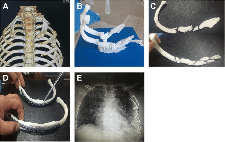
(a) Preoperative scanning revealed long segment comminuted fractures in 3 and 4 ribs. (b) Preoperative 3 and 4 rib models were prepared by 3D printing technology based on CT thin slice scanning. (c), (d) Reduction fracture morphology; the 3D printing model was spliced and the titanium alloy rib locking plate was shaped according to the mode. (e) Postoperative review, the shape of the internal fixator was intact, and the shape of the contralateral rib was perfectly symmetrical compared with preoperative image A
Patient Two
The second patient covered was a 57-year-old male with multiple fractures in his left ribs resulting from a fall. Among these were 4 and 5 costal cartilage and rib 6 anterior costal arch fractures. The surgeons decided to attach the inner end of the titanium plate to the sternum and the outer end of the rib bone due to the nature of the fracture.
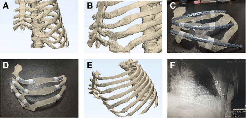
(a) According to the 3D model made by CT, 4 and 5 costal cartilage fractures and 6 costal anterior costal arch fracture can be seen. (b) The 3D model of the fracture end of the rib was adjusted by using 3D software model. (c) The 3D printed 4–6 rib model shows a large gap between the locking plate and the shape of the rib. (d) After giving the shape, the locking plate and the rib paste are in good condition. (e, f) Postoperative review, the shape of the internal fixator was intact, and the shape of the contralateral rib recovered well compared with the preoperative image
Patient Three
This patient is a 64-year-old female who experienced multiple left rib fractures from a traffic accident. Being that the patient is a female, the surgeons attempted to make the operation both minimally invasive and protective of breast tissue. In this operation, the medial side of the locking plate was fixed to the sternal body, and the armpit and sternum were treated through “tunneling open reduction and internal fixation.”
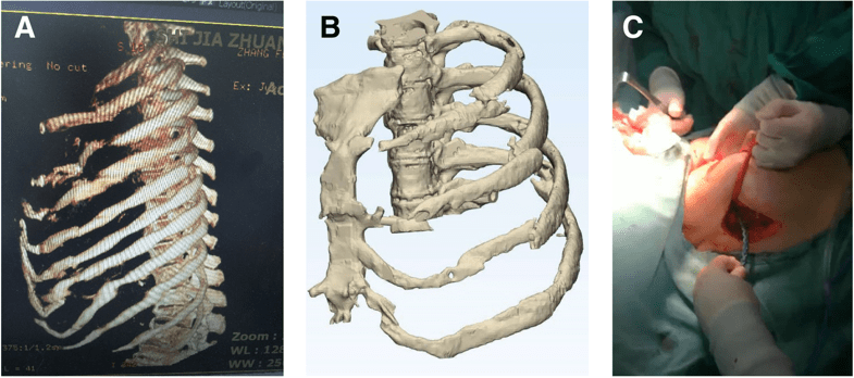
(a) Preoperative scanning revealed 2–11 left rib fractures where 2–6 ribs contained the costal cartilage multiple fractures involving the costal arch. (b) Preoperative 3–5 rib models were prepared by 3D printing technology based on CT thin slice scanning. (c) During the surgery, corresponding pre-shaping locking plates were placed in each rib
Patients Four & Five
Both of these patients were males who experienced multiple rib fractures due to traffic accidents. Being that no breast tissue needed to be preserved, tunneling open reduction and internal fixation was applied. 3D models were utilized in both patients to ensure that the broken ends of the ribs were properly fixated.
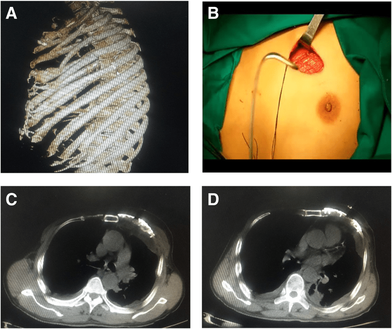
(a) Preoperative scanning revealed 3–6 anterior multiple fractures in the left side involving the cartilage. (b) During the surgery, a 6-cm chest incision was made. (c, d) The postoperative CT locking plate position and thoracic morphology were satisfactory
—
The researchers concluded that preoperative use of 3D printed models can significantly reduce the time spent shaping rib locking plates, as well as the difficulty associated with the treatment of rib fractures. They note that in particularly complex rib fractures, this technique could be beneficial in improving patient outcomes and safety. To better the service patients receive from this technique, the authors call for further clinical verification.
#3dprint : China: Researchers Improve Rib Fracture Surgeries with 3D Printing … In ‘Analysis of the advantages of 3D printing in the surgical treatment of multiple rib fractures: 5 cases report,’ Chinese researchers from Shijiazhuang Third Hospital https://t.co/UpobCFyzjK
— Micron Dental (@microndental) June 25, 2019


 © 2025 Mashup Media, LLC, a Formedics Property. All Rights Reserved.