Immunoglobulin G4-related disease (IgG4-RD) is a multi-system, fibroinflammatory disorder characterized by lymphoplasmacytic infiltration and proliferation. Hypertrophic pachymeningitis is a classic neurologic manifestation of IgG4-RD, which can cause headache, cranial nerve dysfunction, seizures, focal deficits and cognitive decline. Given limited data on this entity, Terrim et al completed a systematic review to better understand the clinical presentation, common findings (laboratory, radiologic, pathologic), treatment outcomes, and prognosis of IgG4-related pachymeningitis [1].
The systematic review involved searching PubMed/MEDLINE, Embase and Scopus databases from inception through May 2023 for case reports and case series of IgG4-related pachymeningitis. Pachymeningitis was defined as presence of thickening and contrast enhancement of the cranial or spinal dura matter, as identified by MRI imaging. ANCA positive cases were excluded. Patient-level data was extracted from articles including neurologic signs and symptoms, systemic involvement, radiologic findings, laboratory findings (serum IgG4 levels, CSF analysis) and pathologic findings. Additional analyses evaluated predictors of outcomes in IgG4-RD pachymeningitis by geography, phenotype of dural involvement (spinal vs cranial), and immunosuppressant treatment received (rituximab vs other).
The study included 148 articles, after excluding irrelevant studies. There were 208 participants, of which 63.5% were male. Most case reports described patients in the United States (21%) followed by Japan (17%) and India (11.5%). The most common presentations were headache and cranial nerve dysfunction, with visual loss, diplopia and/or strabismus the most common type of cranial nerve dysfunction. Seizures were reported in 11% of cases. Other systemic involvement was observed in 49% of cases, most commonly nasal or paranasal (23%), lung (17%), and lymph nodes (15%). Diagnostic neuroimaging often showed preferential involvement of cavernous sinus and middle fossa. Spinal involvement of pachymeningitis was predominantly cervicothoracic.
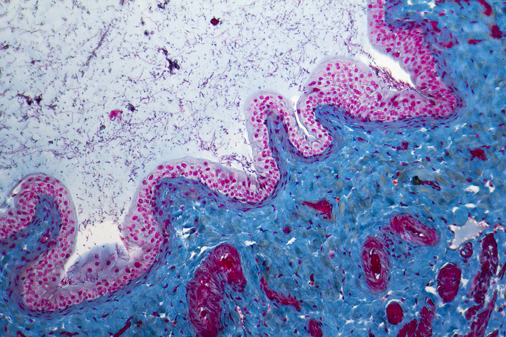
Most patients presented with CSF abnormalities, including pleocytosis in around half (52%) and elevated CSF protein levels in 58%. Serum IgG4 levels were elevated in only 65% of cases. Elevated CSF IgG4 levels were observed in all cases where this information was available. Of patients with documented pathology consistent with IgG4-RD, 81% were meningeal biopsies. While lymphoplasmacytic infiltration and elevated IgG4/IgG ratio were present in almost every biopsy, the storiform fibrosis and obliterating phlebitis were uncommon.
The authors noted mortality was below 1%, though complete clinical or radiologic recovery occurred only in a third of cases and there was a recurrence of disease in 40% of cases. Glucocorticoids were the most commonly used therapy in 85% of patients; rituximab was prescribed as maintenance therapy in 31%. Oral immunosuppressants prescribed included azathioprine (12%), methotrexate (12%), and mycophenolate mofetil (4%). When comparing frequency of refractory disease, Rituximab was associated with lower frequency of refractory disease (9.7%) compared to other immunosuppressants (31%) [p = 0.03], in those with IgG4-RD pachymeningitis with neurologic or systemic biopsy findings suggestive of IgG4-RD.
In conclusion, this systematic review of over 200 cases synthesizes the available evidence on IgG4-RD pachymeningitis. It is important to differentiate from other systemic diseases that can cause pachymeningitis, such as neurosarcoidosis and ANCA-associated vasculitis, and also rule out infectious or neoplastic causes of pachymeningeal enhancement. The study also highlights some diagnostic challenges for IgG4-RD pachymeningitis, including absence of elevated serum IgG4 levels and lack of some of the characteristic pathologic features (storiform fibrosis and obliterative phlebitis) on biopsy in many cases.


 © 2025 Mashup Media, LLC, a Formedics Property. All Rights Reserved.