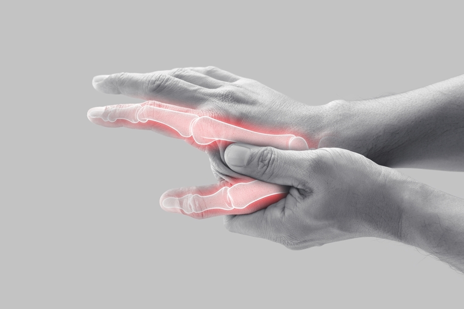
While the relationship between urate deposition and inflammation in gout has been extensively studied, the impact of urate deposition on bone erosion has remained relatively understudied. A recent study, however, has shed light on this important aspect of gout pathology.
The study, published in Frontiers in Endocrinology, aimed to “analyze the influence of tophus in the development of bone destruction and to investigate the relationship between the volume of monosodium urate (MSU) crystals and bone erosion using the improved modified Sharp/van der Heijde (SvdH) erosion scores based on CT images.”
Researchers enrolled 56 patients diagnosed with gout and utilized dual-energy computed tomography (DECT) imaging to measure the volume of MSU crystals in the metatarsophalangeal (MTP) joints. The degree of bone erosion was evaluated using the modified SvdH erosion scoring system based on CT images. Patients were divided into 2 groups: those with urate deposition (UD group) and those without urate deposition (non-UD group). Clinical features and the correlation between erosion scores and urate crystal volume were analyzed.
Among the 560 MTP joints assessed, 80 showed MSU crystal deposition, and 108 showed bone erosion. The study found that patients in the UD group exhibited significantly higher levels of bone erosion compared with those in the non-UD group. The severity of bone erosion was markedly reduced in the non-UD group. Interestingly, both groups had similar levels of serum uric acid, indicating that serum uric acid alone may not be an accurate predictor of the extent of bone erosion. However, the duration of symptoms was significantly longer in the UD group, suggesting a potential association between prolonged urate deposition and increased bone erosion. The UD group also had a higher prevalence of kidney stones, implying a systemic component to the disease.
Furthermore, the study demonstrated a strong positive correlation between the volume of MSU crystals and the degree of bone erosion. This finding suggests that the measurement of MSU crystal volume, in conjunction with serum uric acid levels, can provide valuable insights for optimizing the management of patients with gout. The use of DECT imaging allows for the direct visualization and quantification of MSU crystal deposition, which can contribute to more accurate assessment and monitoring of disease progression.
“To summarize, the correlation between urate volume measured from DECT images and bone erosion were evaluated,” the researchers wrote. “There was no correlation between MSU crystals, bone destruction, and serum uric acid. DECT can effectively monitor MSU deposits and observe changes in bone erosion, and this can be considered as a supplement to serum uric acid measurement in clinical practice to improve the prognostic evaluation of patients with gout and guide follow-up as well as individualized treatment plans.”
Source: Frontiers in Endocrinology


 © 2025 Mashup Media, LLC, a Formedics Property. All Rights Reserved.