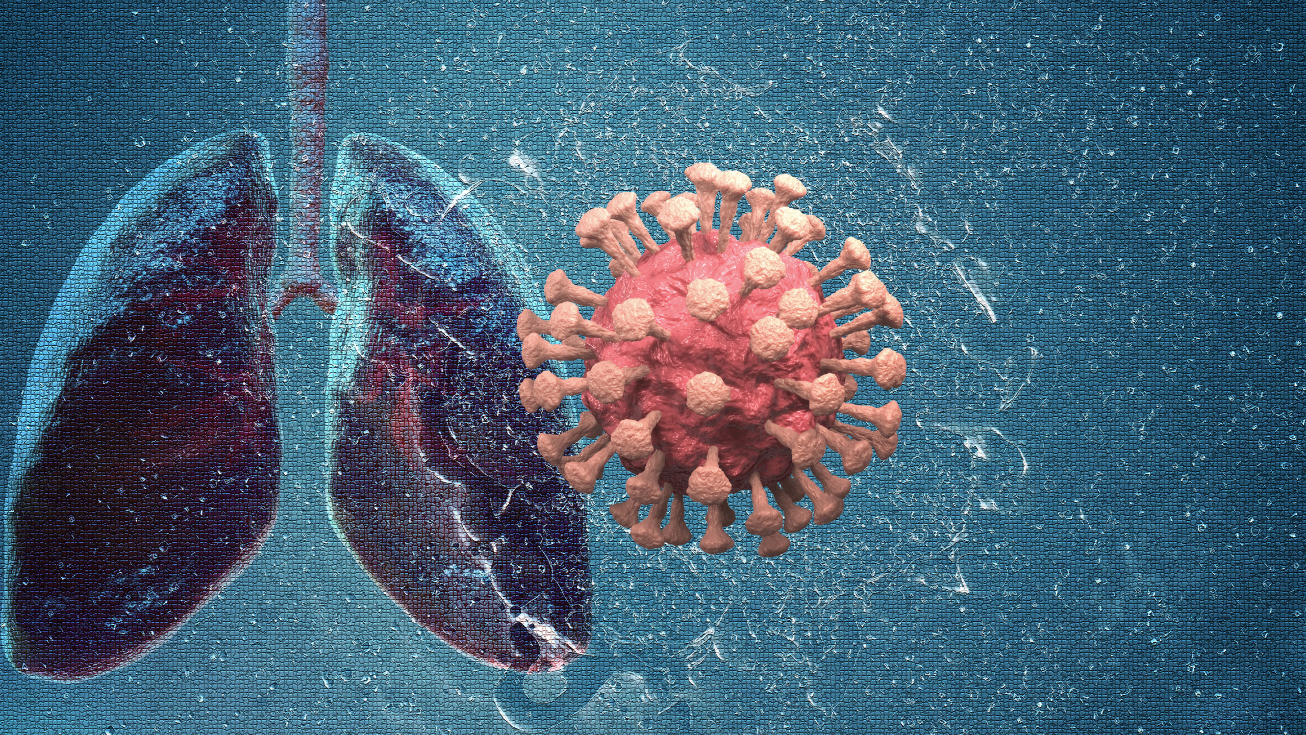
A study presented at the Society of Nuclear Medicine and Molecular Imaging (SNMMI) Annual Meeting shows that 18F-FDG PET/CT imaging is an effective tool to evaluate lung inflammation and monitor the effectiveness of treatment in patients who have residual lung disease and symptoms after COVID-19 infection. The authors said that their study is the first to show that lingering COVID-19 symptoms can be attributed to ongoing lung inflammation.
Respiratory complications such as cough and shortness of breath are common after COVID-19 infection. Many patients have lasting post-COVID lung disease (PCLD) and need oxygen therapy or other treatments, even after negative COVID-19 tests.
“During the pandemic, there was no standard modality to assess residual lung inflammation. It was difficult to gauge the extent and severity of disease even in recovering patients, and hence challenging to start appropriate treatment,” said Yogita Khandelwal, MBBS, a resident doctor at the Sanjay Gandhi Postgraduate Institute of Medical Sciences in Lucknow, Uttar Pradesh, India.
Therefore, the group of researchers conducted a study to evaluate the utility of FDG PET/CT (F-fluorodeoxyglucose positron emission tomography/computed tomography) in assessing the metabolic activity of lung lesions and to evaluate the effect of steroids and antifibrotic drugs.
They enrolled 25 patients (mean age, 56 years; range, 37-71 years; 84% male) who had clinical and image-based evidence of PCLD. All patients had metabolically active lesions in both lungs. On CT, most patients had at least 1 sign of extensive lung disease, such as ground-glass opacities, interstitial thickening, and fibroreticular changes. Of the total sample, 56% has dyspnea during a walk test, 44% had dyspnea at rest, and 52% required oxygen.
The researchers conducted baseline 18F-FDG PET/CT to evaluate inflammation in the lungs. Then patients received steroid and antifibrotic treatment. Follow-up 18F-FDG PET/CT scan was performed after 6 to 12 weeks of therapy to evaluate treatment response and detect remaining fibrosis.
At the time of follow-up, 3 patients had died from respiratory septic shock. The remaining 22 patients had scans a mean 10 weeks after baseline testing. At follow-up, 28% had persistent mild dyspnea during the walk test, no patients had dyspnea at rest, and none required oxygen therapy. Follow-up imaging revealed a significant decrease in the number, size, and FDG-avidity of lung lesions.
The authors concluded that 18F-FDG PET/CT was a sensitive, helpful tool in evaluating the PCLD inflammatory process, monitoring treatment effectiveness, and managing this patient population.


 © 2025 Mashup Media, LLC, a Formedics Property. All Rights Reserved.