A retrospective study discussed clinical and electrophysiological observations of inferior perisylvian epilepsy (IPE). The researchers observed similarities between IPE and mesial temporal lobe epilepsy or rolandic opercular epilepsy. The results of the study were published in Epilepsy & Behavior.
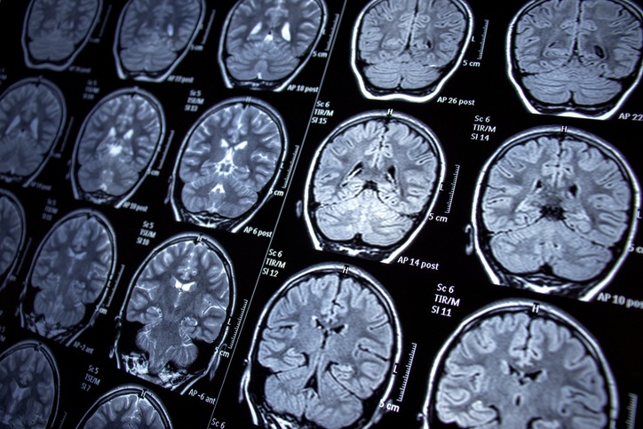
Patients with refractory IPE who underwent resective surgery were retrospectively reviewed. Data analysis included demographic, clinical, neuroelectrophysiological, neuroimaging, surgical, histopathological, and follow-up data. Selected patients underwent quantitative 18F-fluorodeoxyglucose-positron emission tomography (FDG-PET) analysis.
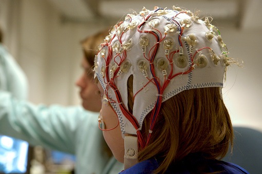
A total of 18 IPE patients were evaluated; among them, “ipsilateral frontotemporal epileptic discharges or its onsets were the dominant interictal or ictal scalp EEG observations,” the study authors reported. The most prevalent documented manifestations were oroalimentary or manual automatism and facial tonic or clonic movements. Upon semiological analysis, patients were stratified into two patterns; in the PET analyses, between-group differences were observed in the neural network.
“Inferior perisylvian epilepsy possesses semiological manifestations similar to those of mesial temporal lobe epilepsy or rolandic opercular epilepsy, hence these conditions should be carefully differentiated. Performing lesionectomy or cortectomy, sparing the mesial temporal structures, was found to be an effective and safe treatment modality for IPE,” the researchers concluded.


 © 2025 Mashup Media, LLC, a Formedics Property. All Rights Reserved.