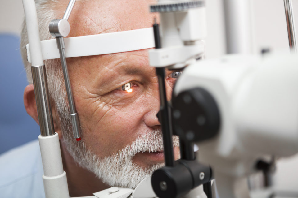
During a simulation workshop on Saturday at AAO 2019, attendees will not only receive a comprehensive review of laser retinopexy, but also practice performing indirect laser retinopexy and laser retinopexy on a simulator.
A laser (light amplification by the stimulated emission of radiation) system is comprised of three parts: an energy source, gain medium, and an optical feedback mechanism. They receive their name from their gain medium. The energy source elevates electrons in the gain medium into a high-energy state, and the energized electron emits an identical photon when light with the appropriate wavelength hits it. This reaction is enhanced and by the optical feedback mechanism, which is normally made up of two mirrors, including one with a small aperture. A small, controlled amount of light exits the system.
Photocoagulation takes place in laser retinopexy. During this thermal process, laser is turned to heat. This results in protein denaturation and creates a scar.
Peripheral retinal breaks include flap tears, operculated holes, atrophic holes, giant retinal tears—all of which could eventually turn into retinal detachment (RD). Risk factors for RD include lattice degermation, meridional folds, enclosed Ora bays, and vitreoretinal tufts. These lesions do not require prophylactic laser retinopexy, but it could be taken into consideration in fellow eyes of patients with RD or other risk factors.
The AAO recommendations for laser use are:
- Flap tear—almost always
- Operculated hole—usually (if symptomatic)
- Atrophic hole—rarely
- Lattice degeneration—rarely (usually in fellow eyes)
The first step in treatment is to find the retinal break. The use of a three-mirror contact lens is recommended. It’s also important to place gently, use lubrication, and view the periphery with different mirrors. When planning the laser, the authors suggest the following:
- Identify all breaks
- Surround each break by three lines of laser burns (about half-width apart)
- Avoid large blood vessels
- Remember to use the mirrors, not the central lens
To begin, use the following laser settings:
- Power: 200 mW
- Time: 0.1 sec
- Area: 200-300 microns diameter
- If the laser does not take, it’s possible that the power is too low, the laser beam is not in focus, or subretinal fluid is present.
Elad Moisseiev, MD, will serve as the course director, and instructors will include Anat Loewenstein, MD; Michaella Goldstein, MD; Lawrence S. Morse, MD, PhD; Glenn C. Yiu, MD; Susanna S. Park, MD, PhD; Jeffrey R. Willis, MD; and Ala Moshiri, MD, PhD.


 © 2025 Mashup Media, LLC, a Formedics Property. All Rights Reserved.