
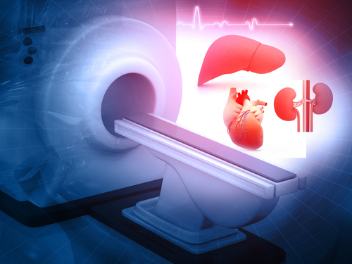
A study compared the association of tumor response evaluated by magnetic resonance imaging (MRI), computed tomography (CT), and ultrasound in patients with hepatocellular carcinoma (HCC) undergoing locoregional treatment (LRT) prior to liver transplantation. The results appeared in Transplantation Proceedings.
In this single-center, retrospective study, researchers analyzed data on 444 patients undergoing liver transplantation for HCC, with a focus on selecting patients who underwent LRT.
According to the results, MRI categorized 60.6% of tumors as nonviable based on the Liver Imaging Reporting and Data System Treatment Response (LR-TR) algorithm, CT categorized 44.4% as nonviable, and ultrasound categorized 62.5% as nonviable. The differences were not clinically significant (P=.779).
The investigators noted the correlation between patients with tumors classified as nonviable by each test and total tumor necrosis in histopathology. Both MRI and ultrasound correctly classified 60% of total tumor necrosis as nonviable, and CT correctly classified 50%. The differences were not clinically significant (P=1).
“Ultrasound and CT have obtained similar results as reevaluation tests to MRI [and] could replace it in case of [the] unavailability of the latter,” the researchers concluded.


 © 2025 Mashup Media, LLC, a Formedics Property. All Rights Reserved.