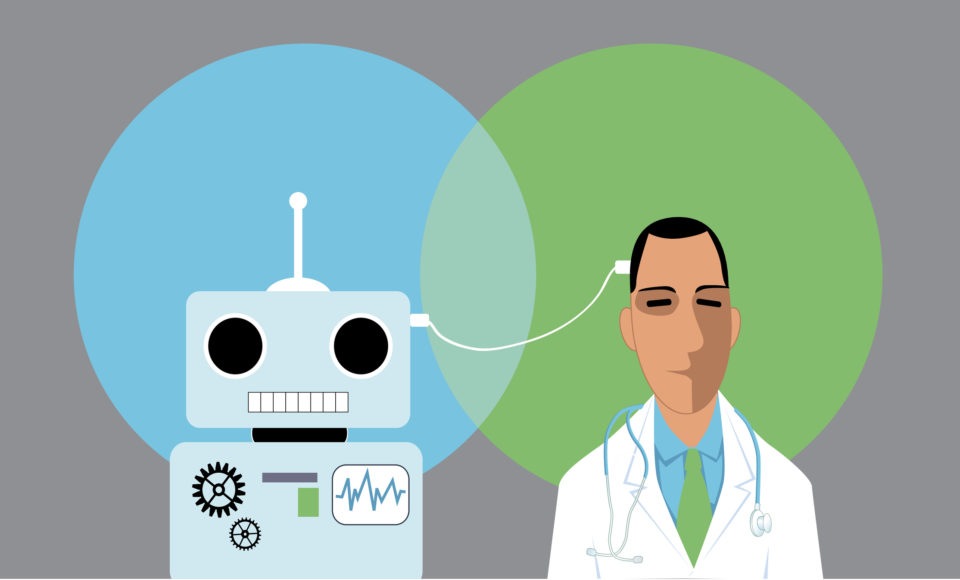
A new study suggests that the results of cardiac magnetic resonance imaging (MRI) scans can be read significantly faster using automated machine learning.
Published in Circulation: Cardiovascular Imaging, the study included 110 patients who underwent scan:rescan cardiovascular magnetic resonance. The researchers identified technique, left ventricular (LV) chamber volumes, LV mass, LV ejection fraction by an expert, a trained junior clinician, and an automated convolutional neural network that trained on nearly 600 (n=599) disease cases. The authors also compared the scan:rescan coefficient of variation and calculated 1,000 bootstrapped 95% confidence intervals using mixed linear effects models.
According to the study results, “clinicians can be confident in detecting a 9% change in LV ejection fraction, with greater than half of coefficient of variation attributable to intraobserver variation.” Scan:rescan precision was similar between expert, trained junior clinician, and automated observers. However, the most interesting finding was that the automated analysis was 186x faster than the human readers (0.07 minutes versus 13 minutes).
“Automated machine learning analysis is faster with similar precision to the most precise human techniques, even when challenged with real-world scan:rescan data,” the researchers wrote in their study. “Assessment of multicenter, multi-vendor, multi-field strength scan:rescan data permits a generalizable assessment of machine learning precision and may facilitate direct translation of machine learning to clinical practice.”
The added in their clinical perspective that “[a]utomated cardiovascular magnetic resonance analysis should, therefore, be adopted globally to gain from time-saving and standardization benefits.”
Study authors Charlotte Manisty, MD, PhD, said that the results are encouraging, and can envision scenarios where artificial intelligence helps clinicians perform “super human” tasks.
“Cardiovascular MRI offers unparalleled image quality for assessing heart structure and function; however, current manual analysis remains basic and outdated. Automated machine learning techniques offer the potential to change this and radically improve efficiency, and we look forward to further research that could validate its superiority to human analysis,” Dr. Manisty said in a press release. “Our dataset of patients with a range of heart diseases who received scans enabled us to demonstrate that the greatest sources of measurement error arise from human factors. This indicates that automated techniques are at least as good as humans, with the potential soon to be ‘super-human’—transforming clinical and research measurement precision.”
Only 186X faster? 😉#AI automated heart MRI analysis@mrimypacemaker @UCL_ICS Charlotte Manisty and colleagueshttps://t.co/ie6wjHsBXo @CircAHA #openaccess pic.twitter.com/5PpCuVFOQ7
— Eric Topol (@EricTopol) September 24, 2019
— Sumanth Raman (@sumanthraman) September 24, 2019



 © 2025 Mashup Media, LLC, a Formedics Property. All Rights Reserved.